
Forgot your password?
A Beginner’s Guide to Mitral Transcatheter Edge-to-Edge Repair (mTEER): Patient Selection, Procedural Fundamentals and Clinical Evidence
Dr Ahmed Salem, Interventional and Structural Cardiology Fellow, Leeds Teaching Hospitals NHS Trust
TRANSCATHETER EDGE-TO-EDGE REPAIR IN MITRAL REGURGITATION
PART 1 — Pre-procedure Assessment and Planning
Mitral regurgitation (MR) is the most prevalent valvular heart disease in the developed world, affecting approximately 2% of the general population and rising to over 10% in those aged over 75 years.1
Untreated severe MR leads to progressive left ventricular (LV) remodelling, heart failure, and premature death.2 Surgical mitral valve repair or replacement has historically been the definitive treatment; however, a significant proportion of patients are deemed inoperable or at prohibitively high surgical risk due to advanced age, frailty, or comorbidities.3 Mitral transcatheter edge-to-edge repair (M-TEER) has emerged as a safe and effective alternative. MitraClip (Abbott Vascular) remains the best-established device platform globally, with over 200,000 procedures performed to date, while PASCAL (Edwards Lifesciences) is increasingly used as an alternative M-TEER system in contemporary practice.4 Understanding the fundamentals of patient selection and pre-procedural planning is essential for any cardiologist involved in the care of patients with valvular heart disease.
Understanding Mitral Regurgitation: Primary versus Secondary
A critical first step in evaluating any patient for TEER is establishing the mechanism of MR, as this determines both the patient pathway and the expected benefit from intervention.
Primary MR (also termed degenerative MR) arises from structural abnormality of the mitral valve apparatus itself — including leaflet prolapse, flail leaflet, or chordal rupture (Figure 1), most commonly due to myxomatous degeneration.5 In these patients, the mitral valve is the primary pathology, and effective repair restores normal haemodynamics.
Secondary MR (also termed functional MR) arises from LV dysfunction or dilatation causing annular dilatation and leaflet tethering, in the absence of intrinsic valve pathology.5 Here, the mitral valve is structurally normal but geometrically distorted. Secondary MR is most commonly encountered in patients with heart failure with reduced ejection fraction (HFrEF), where it perpetuates a vicious cycle of further LV remodelling and worsening haemodynamics. The distinction carries important prognostic and therapeutic implications, and underpins the differing evidence bases for TEER across the two aetiologies, as discussed in Part 2.
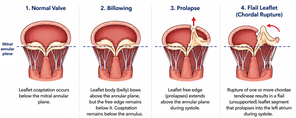
Figure 1:Schematic illustration demonstrating the anatomical progression from a normal mitral valve to leaflet billowing, prolapse and flail leaflet secondary to chordal rupture.
Patient Selection
Optimal patient selection is fundamental to a successful outcome and is best achieved through a dedicated valvular heart disease multidisciplinary team (MDT), comprising interventional cardiologists, cardiac surgeons, imaging specialists, heart failure physicians, and specialist nurses (Figure 2).
In patients with primary MR, TEER is indicated in those with severe symptomatic MR who are deemed inoperable or at high surgical risk, and in whom valve anatomy is suitable.6 Surgical repair remains the gold standard in operable patients with favourable anatomy, given its superior long-term durability. However, in centres with high surgical expertise, TEER may be considered in selected low-risk patients, particularly where anatomy is highly suitable and patient preference favours a less invasive approach.
In patients with secondary MR, the threshold for intervention is guided not only by MR severity but by the degree to which MR is felt to be contributing to symptoms and LV dysfunction — the concept of “proportionate” versus “disproportionate” MR.7 TEER is indicated in patients with severe secondary MR who remain symptomatic despite optimal guideline-directed medical therapy (GDMT), including maximally tolerated doses of neurohormonal blockade and cardiac resynchronisation therapy where appropriate.6
A third and increasingly recognised entity is atrial functional MR, which arises from left atrial dilatation and annular enlargement in the context of atrial fibrillation or heart failure with preserved ejection fraction, in the absence of significant LV dysfunction or intrinsic leaflet pathology. The evidence base for M-TEER in this group is less mature than for degenerative or ventricular secondary MR, and careful MDT discussion — including rhythm strategy, rate control, and anticoagulation — is essential before considering intervention.
Across all aetiologies, careful assessment of frailty and comorbidity burden is essential. As with TAVI, frailty confers a higher risk of poor outcomes and reduced quality-of-life benefit following intervention,8 and patients with a high frailty index or limited life expectancy due to non-cardiac comorbidities are unlikely to derive meaningful benefit from TEER.
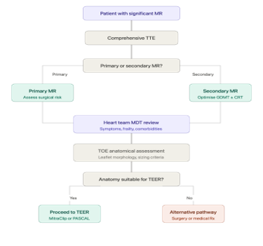
Figure 2: Overview of the TEER patient pathway. Simplified algorithm demonstrating assessment, multidisciplinary discussion and anatomical evaluation prior to mTEER
Pre-procedure Anatomical Assessment
All patients being considered for TEER require comprehensive multi-modality imaging to confirm anatomical suitability, guide device selection, and anticipate potential procedural challenges.
Transthoracic echocardiography (TTE) is the first-line imaging modality and provides essential information regarding MR mechanism and severity, LV dimensions and systolic function, and mitral valve morphology. Grading of MR severity should be multiparametric, integrating qualitative, semi-quantitative, and quantitative measures in accordance with current guidelines.5,6
Transoesophageal echocardiography (TOE) is indispensable in pre-procedural planning and provides superior resolution of mitral valve anatomy. Key measurements guiding TEER eligibility include mitral valve area (MVA ≥4.0 cm² to accommodate the device without inducing iatrogenic mitral stenosis), posterior leaflet length (≥10mm), flail gap (<10mm) and flail width (<15mm) in primary MR, and coaptation depth (<11mm) and coaptation length (≥2mm) in secondary MR.9 The presence of severe mitral annular calcification, rheumatic valve disease, or significant leaflet cleft may preclude TEER and should be systematically evaluated. Unlike PCI, TEER is fundamentally an imaging-guided procedure. Close communication between the interventional cardiologist and echocardiographer throughout the case is essential and is often a defining feature of procedural success.
Computed tomography (CT) plays a complementary role, particularly in assessment of the interatrial septum to guide transseptal puncture planning, and in providing precise annular dimensions where TOE data is inconclusive. CT is also valuable in identifying anatomical variants or co-existing pathologies that may influence procedural strategy.
Conclusion to Part 1
TEER represents a major advance in the management of mitral regurgitation in patients unsuitable for surgery. Careful pre-procedural assessment — encompassing clinical evaluation, MDT discussion, and multi-modality imaging — is the cornerstone of patient selection and procedural success. Part 2 will describe the procedural steps, evidence base, and potential complications of TEER.
PART 2 — The Procedure, Evidence, and Complications
The Procedure
TEER is performed in a dedicated catheterisation laboratory or hybrid theatre under general anaesthesia, with continuous TOE guidance throughout. The procedure replicates the surgical Alfieri stitch technique, whereby the anterior and posterior mitral leaflets are approximated at the point of maximum regurgitation, creating a double-orifice valve and reducing MR.10
Vascular access is obtained via the right femoral vein with the aim to do a trans-septal puncture. For the beginner, trans-septal puncture is arguably the single most important step of the procedure. An optimally positioned puncture greatly facilitates device steering and leaflet grasping, whereas a poorly positioned puncture is difficult to rescue later in the case. Under fluoroscopic and TOE guidance, the puncture site should be aimed at the posterosuperior aspect of the interatrial septum, typically 3.5–4.5cm above the mitral annular plane.11 Adequate trans-septal height is critical — too low a puncture compromises manoeuvrability of the delivery system within the left atrium and makes precise leaflet grasping difficult (Figure 3).
Following trans-septal puncture, the steerable guide catheter and clip delivery system are advanced into the left atrium. Under live three-dimensional TOE guidance, the M-TEER device is steered above the mitral valve orifice and oriented perpendicular to the line of coaptation. The device is then advanced into the left ventricle, pulled back to grasp both leaflets simultaneously, and partially closed to assess leaflet insertion and resulting MR reduction. If satisfactory — defined as residual MR of mild or less with an acceptable transmitral gradient — the device is fully closed and detached. Additional devices may be deployed if residual MR remains significant, provided the transmitral gradient remains acceptable (mean gradient <5mmHg).9 Real-time three-dimensional TOE provides dynamic assessment of valve pathology, MR severity, device positioning, and procedural result throughout each stage of the procedure (Figure 4).
The PASCAL device (Edwards Lifesciences) operates on the same edge-to-edge principle but incorporates independent leaflet clasps and a central spacer element, which may confer advantages in certain anatomical configurations such as large flail segments or restricted leaflet mobility.12 It is increasingly used alongside MitraClip across UK structural centres.

Figure 3: Transoesophageal echocardiographic image demonstrating optimal transseptal puncture height during M-TEER. The red line illustrates a puncture height of 4 cm above the mitral annular plane, with visible tenting of the interatrial septum confirming appropriate needle position at the posterosuperior aspect of the fossa ovalis. Adequate puncture height is critical to ensure manoeuvrability of the delivery system within the left atrium and facilitate precise leaflet grasping.
Figure 4: Transoesophageal echocardiographic assessment during M-TEER. Video 1: Three-dimensional TOE demonstrating mitral valve pathology (P2 prolapse) prior to intervention. Video 2: Three-dimensional TOE with colour Doppler showing the mitral regurgitation jet. Video 3: Post-procedural colour Doppler confirming the double-orifice configuration following clip deployment. Video 4: Three-dimensional TOE with colour Doppler demonstrating reduction in mitral regurgitation following successful de
Evidence Base
The pivotal EVEREST II trial established the safety and feasibility of MitraClip in primary MR, demonstrating non-inferiority to surgery for the composite endpoint of freedom from death, surgery, and grade 3+ MR at one year, with a significantly lower rate of major adverse events in the TEER arm.13 Longer-term follow-up confirmed durable clinical benefit, though surgical repair remained superior in terms of MR reduction in anatomically suitable patients.
The landscape for secondary MR was defined by two landmark trials published simultaneously in 2018. The COAPT trial demonstrated that MitraClip in addition to GDMT significantly reduced hospitalisation for heart failure and all-cause mortality at two years compared to GDMT alone in carefully selected patients with disproportionate secondary MR.14 In contrast, the MITRA-FR trial showed no benefit of TEER over GDMT alone.15 The divergence between these trials is largely attributed to differences in patient selection — COAPT enrolled patients with more severe and disproportionate MR relative to LV volumes, reinforcing the importance of the proportionality concept in identifying patients most likely to benefit.7 More recently, the RESHAPE-HF2 trial independently confirmed a significant reduction in heart failure hospitalisations with M-TEER in patients with symptomatic secondary MR despite GDMT, further strengthening the evidence base for carefully selected patients. These findings are reflected in current ESC guidelines, which recommend TEER in symptomatic patients with severe secondary MR despite GDMT, provided anatomy is suitable and the Heart Team considers the patient unlikely to benefit from surgery.6
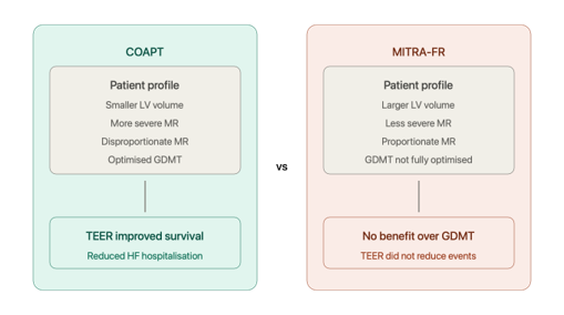
Figure 5: Why COAPT succeeded whereas MITRA-FR did not.
Conceptual illustration of proportionate versus disproportionate secondary mitral regurgitation.
Complications
As with any structural intervention, TEER carries a defined complication profile that should be discussed openly with patients during the consent process (Figure 6).
Single leaflet device attachment (SLDA) occurs when the clip engages only one leaflet, either intra-procedurally or following detachment, and represents the most important device-specific complication with an incidence of approximately 1–3%.16 It may result in acute worsening of MR and occasionally necessitates emergency surgery. Careful TOE assessment of leaflet insertion prior to clip detachment is the principal safeguard.
Iatrogenic mitral stenosis can result from excessive leaflet tissue capture or deployment of multiple clips, leading to a reduction in effective mitral valve area. A post-procedural mean transmitral gradient exceeding 5mmHg should prompt reassessment of clip configuration.9
Residual or recurrent MR may occur acutely due to incomplete clip closure or suboptimal positioning, or in the longer term due to clip detachment or disease progression. Careful intra-procedural TOE assessment minimises acute residual MR, though some degree of mild residual MR is not uncommon and is generally well tolerated.
Trans-septal complications including cardiac tamponade are rare but recognised, with an incidence of less than 1% in contemporary series.17 Meticulous technique and continuous TOE guidance during puncture are essential mitigates.
Vascular access complications are less frequent than in arterial structural procedures given the venous access route, but haematoma, arteriovenous fistula, and rarely deep vein thrombosis may occur.

Figure 6: Cartoon illustration of the major procedural and device-related complications of TEER, highlighting the importance of meticulous transseptal puncture, adequate leaflet grasping, and careful assessment of residual mitral regurgitation and transmitral gradients before device deployment.
Conclusion
M-TEER is a safe, effective, and increasingly accessible treatment for patients with significant MR who are unsuitable for or decline surgery. Procedural success is underpinned by meticulous patient selection, rigorous anatomical assessment, and experienced TOE guidance throughout. As the evidence base matures and device technology continues to evolve, TEER is likely to expand further across the spectrum of MR severity and aetiology. A working knowledge of the procedure and its evidence base is therefore of value to all cardiologists managing patients with valvular heart disease.
Acknowledgements
The authors wish to thank Dr Jaydeep Sarma for his supervision and expert guidance in the preparation of this manuscript, and to Dr William Smith and Dr Mike Sosin for kindly providing the echocardiographic materials used in Figure 4.
References: